Exploring Radiography: A Window into Modern Medical Imaging
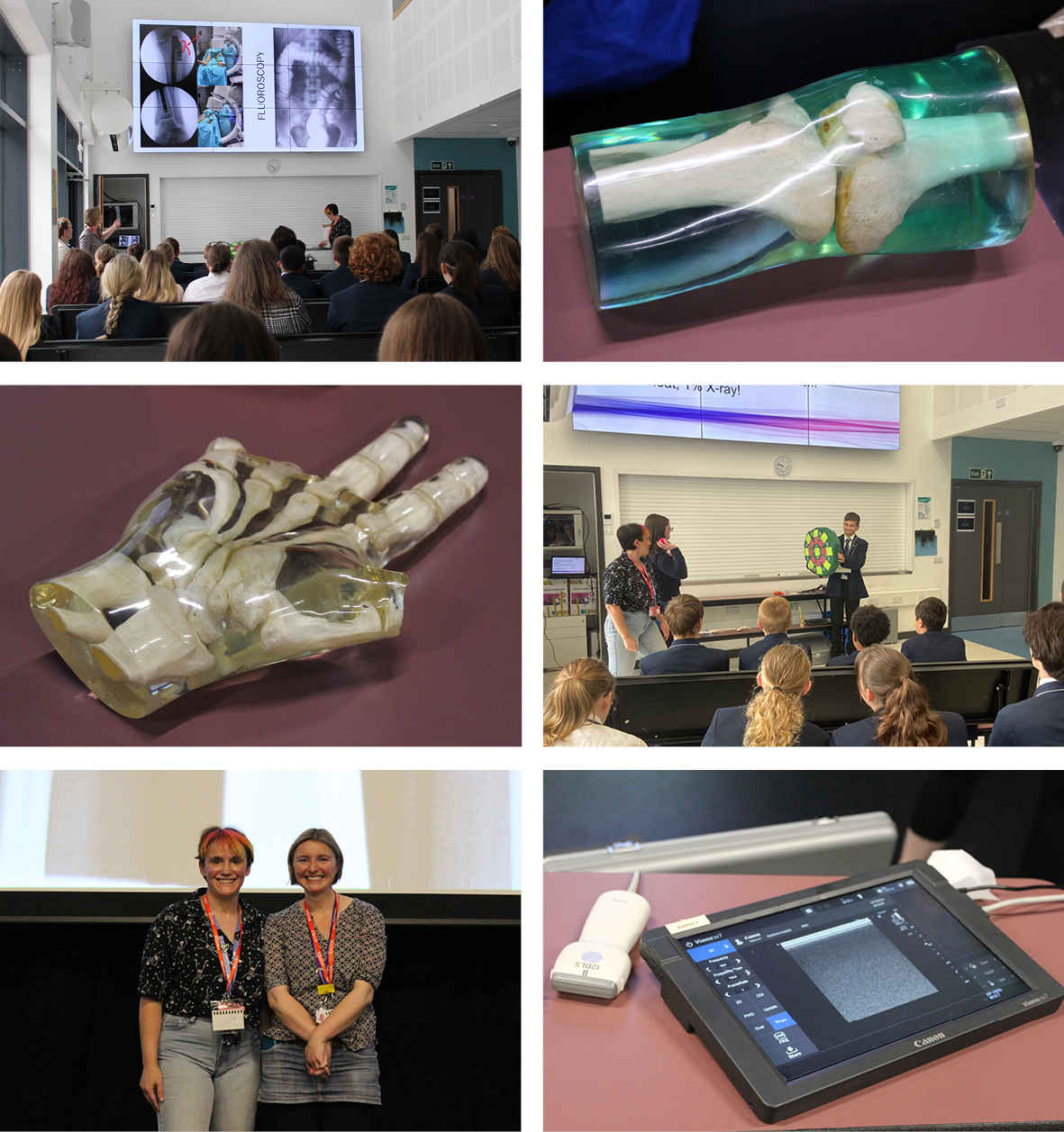
We were delighted to welcome Janet Villars and Lisa Hurndall for an engaging and inspiring session exploring the world of radiography. Their visit gave students fascinating insights into the role of a radiographer, the science behind X-rays and ultrasounds, and how medical imaging technology continues to evolve.
The session began with a brief history lesson, taking students back to 1895 when German scientist Wilhelm Conrad Röntgen accidentally discovered X-rays. He famously captured the first X-ray image of his wife's hand — a moment that transformed medical diagnostics forever.
Students were intrigued to learn that radiation is part of everyday life. From eating bananas to walking across Dartmoor’s granite terrain, we regularly encounter low levels of radiation. This real-world context helped students understand how natural and medical radiation coexist.
A highlight of the session was an interactive quiz where students examined a range of medical images — including mammograms, fractured jawbones, ultrasound scans of twins, and even shotgun pellets embedded in a knee. This hands-on approach brought learning to life and sparked curiosity about careers in radiography.
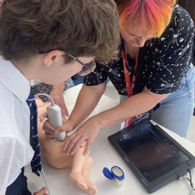
Janet shared her passion for her work with the NHS and her role at a private IVF clinic, offering students a real-world view of the diverse career paths available in healthcare. When asked by Year 9 student Bella why patients need a full bladder for an ultrasound scan, Janet explained: “A full bladder improves visualisation of organs like the uterus and kidneys, enhances image clarity by transmitting sound waves more effectively, and assists in diagnosing conditions such as UTIs or bladder stones.”
Lisa brought the science of radiography to life through interactive demonstrations. Students acted out how X-rays work, discovering that only 1% of the energy used actually creates the image — the rest becomes heat. They also mimicked how ultrasound machines operate, learning how vibrating crystals send sound waves through the body, which then bounce back to form detailed images.
Katie in Year 9 reflected, “It was so inspirational — I learnt so much new knowledge today.”
Students were particularly impressed by the advances in imaging technology. Lisa explained how modern scans can now replace traditional autopsies, offering vital insights for families whose religious beliefs prohibit invasive procedures.
Feedback from students across year groups was overwhelmingly positive:
-
Year 10 student Soheil said, "It was great when we got to hold items and take part in demonstrations. Seeing how ultrasound and X-rays work made it all so clear."
-
Year 12 student Ife commented, "I loved the interactive elements — learning by doing really helped me understand how these technologies work."
-
Tess, in Year 10, added, "They were so knowledgeable but explained everything in a way we could understand. It’s definitely a career I’m now considering."
Mrs Chalmers expressed her sincere thanks to Janet and Lisa for their time, enthusiasm, and the brilliant way they shared their expertise. The session not only educated but also inspired many students to consider future careers in radiography and healthcare.